By Thomas “Tad” Riley
B.S. Physics, Class of 2016
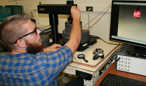
 I worked this summer in Dr. Arthur Smith’s lab with our Atomic Force Microscope (AFM) system that was recently refurbished by Anfatec, a small family-owned company in Germany. I tested all the functionality of the microscope, developing written procedures to operate the AFM, and I’m using it this semester to study materials synthesized in our molecular beam epitaxy (MBE) growth facility.
Samples I examined include a standard test sample (grating) provided by the company, and after that some thin film samples such as manganese nitride grown on magnesium oxide substrates and other magnetic materials grown in our MBE system. The AFM is capable of acquiring both magnetic as well as topographical information about the samples of interest. It is highly sensitive with spatial resolution down to just a few nanometers or less.
Last spring, Anfatec President Dr. Anne-Dorothea Müller traveled from Germany to pay us a visit, bringing with her an updated controller unit. I spent a day and a half working directly with Dr. Müller, and learning all about the functionality of the system.
Working one-on-one with Dr. Müller was a great experience. I was able to rapidly learn what the AFM system is capable of and how it operates in relation to the software. This afforded me the knowledge and skillset to take several hundred images by the end of summer. It was great getting to experience firsthand what it is like to have international collaboration between scientists in private industry and in government-funded research.
This fall I’m fortunate enough to still be working with Dr. Smith. I’m continuing to take magnetic and topographical images of samples grown by several different members in Dr. Smith’s lab group. A main focus is trying to prove that the images taken are true magnetic images rather than images influenced by surface features. I’m also working with a partner to produce simulated magnetic images. This will help to both validate magnetic force microscopy theory and to provide a comparative reference for images taken.
Tad Riley – Intern with Dr. Arthur Smith – junior at Ohio University – College of Arts & Sciences – physics major


Comments